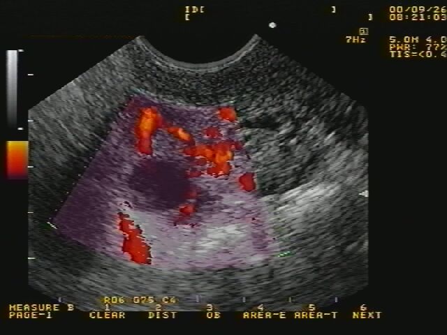
Fig.1: Original doppler ultrasound image
Ovary contains other structures like nerve fibers, lymphatic glands and blood vessels. This quantity of blood, and thus the number and the size of blood vessels around the dominant follicle, is very interesting for physicians, because it can reveal an abnormal situation in the ovary. It can even be predictive of a subsequent follicular response in an In Vitro Fertilization program, because it may be useful in determining the appropriate dose of gonadotrophin to administer in order to achieve optimal response. Indeed the dominant follicle is supposed to be the most fed by blood, and too much or too less quantity of blood around the dominant follicle can trouble its normal development.
At this time, there is no known way to automatically compute the quantity of blood around the follicle. Physicians are able to visualize it, and to know the flow of blood or the resilience in the vessels, owing doppler images. But they are unable to estimate this quantity of blood around the follicle, that is a very important parameter for them.
This project has been realised in collaboration with Prof. Dr. Vlaisavljević, who is gynecologist at the Teaching hospital of Maribor.

Fig.1: Original doppler ultrasound image
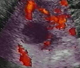
Fig. 2: Extracted subimage for further processing
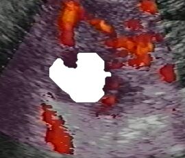
Fig. 3: Detected follicle using cellular neural networks
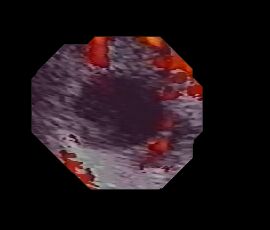
Fig. 4: Condition image (useful part: 5mm around follicle)
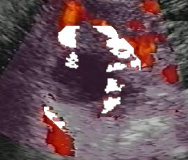
Fig. 5: Blood vessels in 5 mm pass around follicle
After processing the entire image sequence (usually around 500 doppler images) the following quantitative results are obtained:
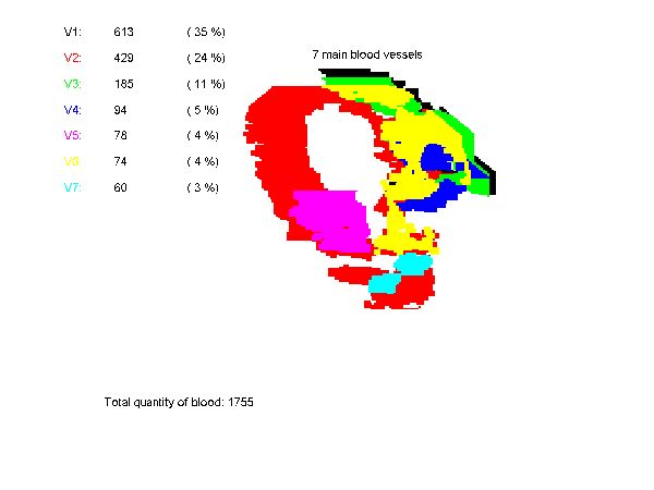
Fig. 6: Importance and location of the blood vessels in the follicle
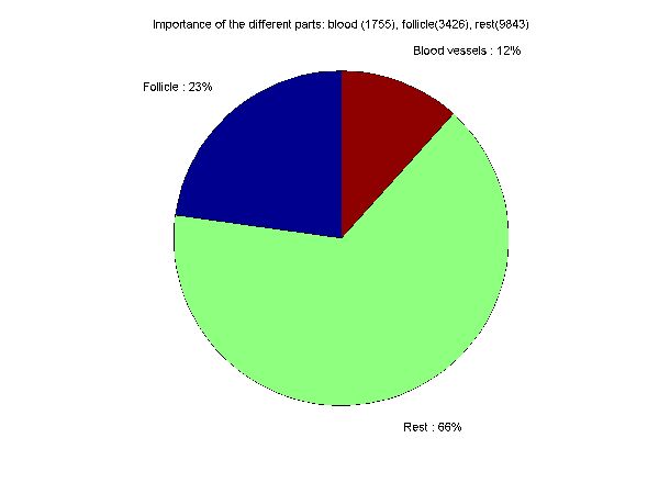
Fig. 7: Relative importance and quantitative values of the different elements
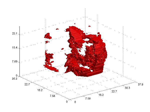
Fig. 8: 3D reconstruction of blood vessels in 5 mm pass around
follicle (unit of measure is mm)
N. Sergent and D. Zazula, "Quantitative and automated estimation of blood around the follicle in the ovary", Technical report, University of Maribor, Faculty of EE and CS, Maribor, 2000.