|
|
|
|
|||||
|
Technical Progress
|
TECHNICAL PROGRESS - SUMMARY OF ACTIVITIES
1.
Acquire high-quality static Magnetic Resonance (MR) images of normal and
pathological knees.
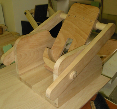
TECHNICAL PROGRESS - MR COMPLIANT EXERCISE RIG The purpose of the MR compliant exercise rig is to permit the validation of the quality of the fine element (FE) models created using the SimBio environment. The rig must allow a volunteer or patient to undertake a controlled exercise protocol while exerting known light forces and the rig must be fully MR compliant, that is, it must not utilise any metal or ferro-magnetic materials in its construction. The MR exercise rig is required for acquisition of pseudo-dynamic in-vivo MR images. Partner SBM is reproducing MR compliant exercise rig with some minor modification according to a scheme provided by the partner USFD, while partner UNI-MB is developing optical sensor for measuring a force on a pedal of rig.
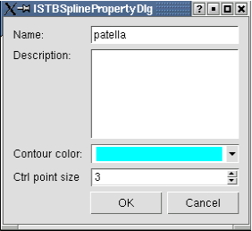
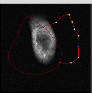
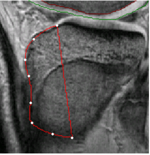
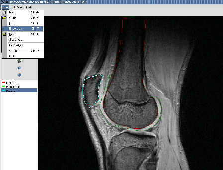
TECHNICAL PROGRESS - AN IST TOOL An Image Segmentation Tool (IST) application has been designed as a MR image manipulation and analysis software. This tool is in the first place meant as a helping tool for manual segmentation/annotation of the MR images (not necessarily knee images). It enables that clinicians manually annotate (segment) every 2D slice of the MR sequence. Marked interesting regions for each 2D slice are afterwards stored as a XML document (output). All
features that are supported by the IST application are as follows: A segmentation tool, a core of IST application, has been added as utility for a verification phase inside the SimBio project, but it can be used also for other intentions. It enables to create, manipulate, delete and save contours or regions. It has advanced GUI (e.g. popup menus). Each annotation (e.g. meniscus) is represented with own colour, some descriptive text etc. There are integrated some user friendly routines like copying annotations from previous slices, importing annotations, zoom, etc.
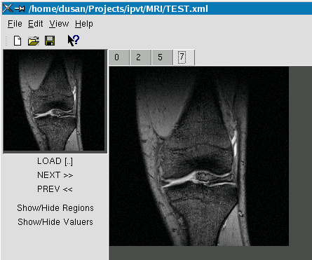
TECHNICAL PROGRESS - AN IPVT TOOL Another auxiliary tool being developed in Maribor is a tool for validating SimBio image segmentation results. An Image Processing Verification Tool (IPVT), as this tool is named, is meant as a tool for statistical validation of the SimBio environment image processing results. The IPVT tool statistically evaluates segmentation ability of the SimBio environment registration routines based on comparison with references provided by clinicians.
|
||||||||||||||||||||||||