

Objective and accurate assessment of skeletal muscle excitation patterns and their interactions after stroke
(Scientific collaboration between the Republic of Slovenia and United States of America from Jan. 1, 2018 to Dec. 31, 2019)
Stroke routinely affects the sensorimotor systems of the brain, seriously hindering the quality of life of survivors. Rehabilitation is very time-consuming, costly, and frequently frustrating for a stroke survivor. Muscle excitation patterns after stroke or during its rehabilitation are not well understood, mainly due to significant methodological limitations in in-vivo assessment.
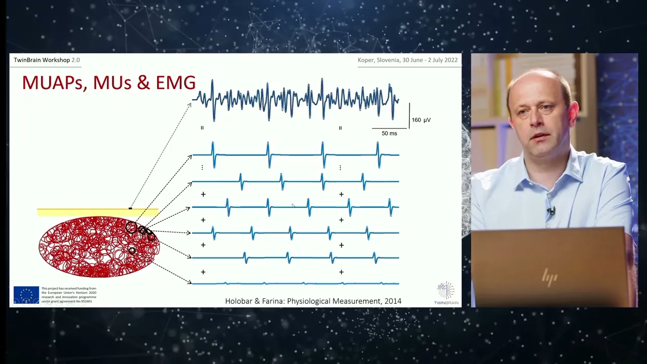
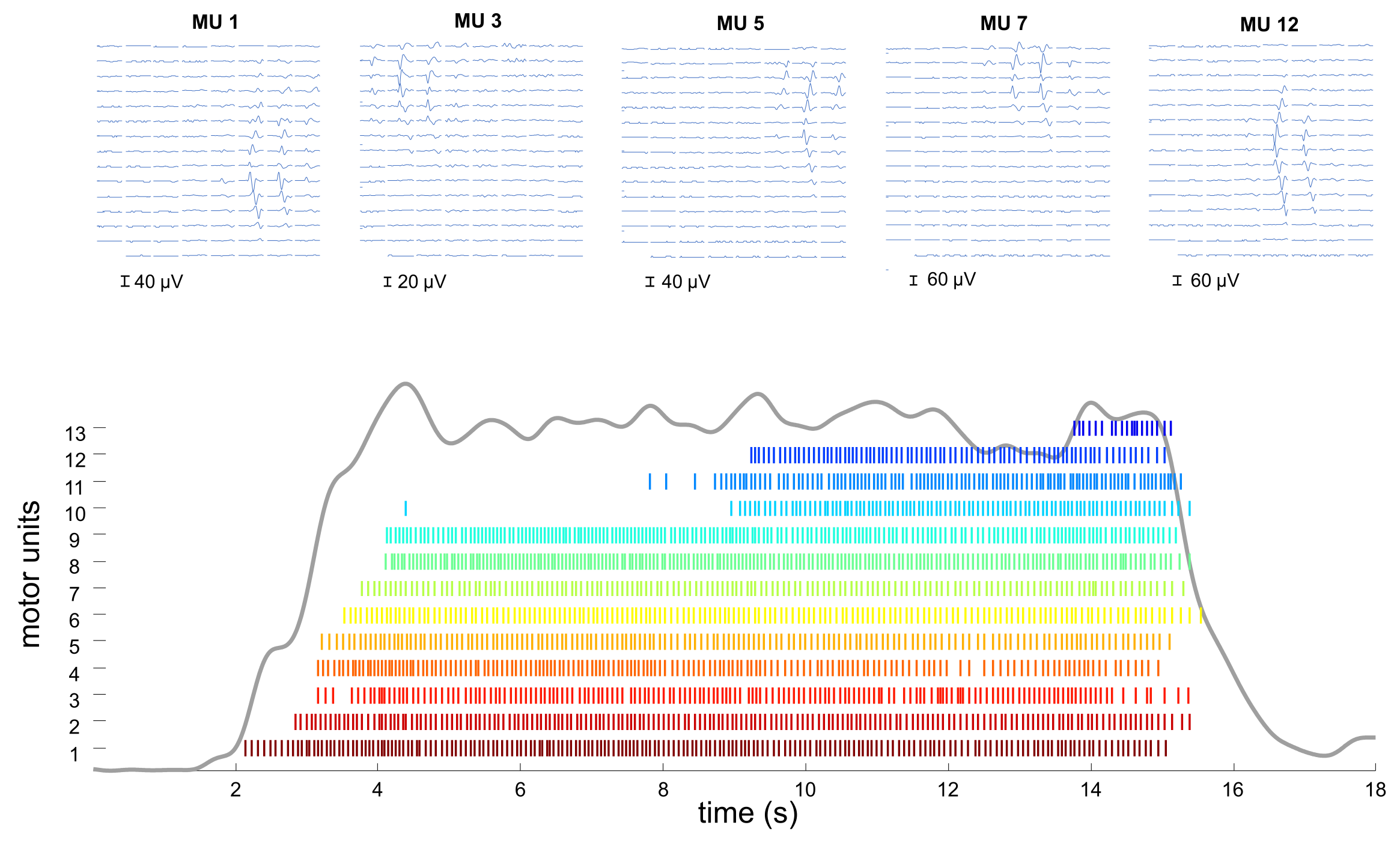
In skeletal muscles, hundreds of muscle fibers are usually innervated by the same motor neuron (MN), forming what is referred to as a motor unit (MU). The Central Nervous System (CNS) modulates active muscle force by modulating the number of recruited MUs and their firing rates. In healthy conditions, several MNs receive common or at least similar supra spinal excitation patterns, dictating common modulations of innervated MU firing rates. MNs also exhibit individual firing rate oscillations, resulting from variations in synaptic inputs (so-called synaptic noise) and MN excitability.
Stroke has been demonstrated to cause significant alterations in MU recruitment and firing patterns. In particular, MU firing rates routinely decrease, whereas their variabilities increase, demonstrating incapability of a subject to precisely control active force generation. The pathophysiology behind these mechanisms is not well understood. In particular, the level of common oscillations of MNs in the same muscle has never been systematically quantified. Analysis of interactions among synergistic and antagonistic muscles is also lacking, hindering the detailed assessment of impairments in each individual stroke subject and their alterations due to various therapies.
In past decades, the intramuscular electromyogram (EMG) became the de facto standard for muscle analysis, building on extensive and long-lasting knowledge of structural changes in MUs due to different pathologies. However, intramuscular EMG recordings are highly selective, limiting detection of electrical activity to muscle fibers that are less than 2mm away from the needle. As a result, only a few concurrently active MUs are routinely recorded by a single needle electrode. Simultaneous recordings from different needles are impractical and are not established in clinical practice. Due to these limitations, standard clinical EMG analysis has focused on the analysis of MU action potential (MUAP) morphology, whereas the analysis of common and individual MU/MN firing patterns has received less attention. Yet, MU firing patterns code each and every muscle movement in humans.
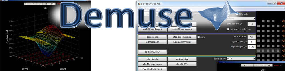
The aforementioned methodological limitations have been recently mitigated by multichannel surface electrode recordings. Surface electrodes are much less selective than the needles, and detect contributions from several tens of MUs. This results in a highly complex signal that has only recently been successfully decomposed into contributions of individual MUs, using computer algorithms that have been pioneered by our group. This methodology supports detailed MU investigations, even at maximum contraction levels. Both common and individual variations of MU firing rates can be quantified by analyzing the firing patterns of several concurrently active MUs. The higher the number of analyzed MUs, the higher the accuracy of muscle excitation assessment. We have recently demonstrated identification of up to 70 concurrently active MUs in a muscle.
In this bilateral project, the aforementioned multichannel surface EMG methodology developed at the University of Maribor has been applied to investigate the main characteristics of muscle excitations in hemiparetic stroke subjects, recruited by Shirley Ryan AbilityLab. In particular, we:
- Studied the feasibility of proposed muscle assessment methodology for long-term objective monitoring of the neuromuscular system during rehabilitation.
- Utilized the proposed muscle assessment to characterize alterations in MN/MU properties subsequent to prescribed interventions for specific stroke related motor impairments, in particular botulinum toxin injections for the reduction of spasticity in muscle.
- Identified and analyzed common features of MN/MU firing patterns in individual muscles of hemiparetic patients.
- Estimated the time course of synaptic excitation in motor neurons innervating spastic muscles of stroke survivors.
- Characterized the spatial properties of muscle activation in stroke impaired muscles.
These studies will provide novel knowledge about the stroke pathophysiology and its effect on the sensorimotor system.
Projektni partnerji
Laboratorij za sistemsko programsko opremo,
Faculty of Electrical Engineering and Computer Science,
Univerza v Maribor
Principal Investigator in Slovenia: Prof. Aleš Holobar
Shirley Ryan AbilityLab
Chicago, IL, USA
Principal Investigators in USA:
Prof. William Zev Rymer, MD, PhD,
Nina L Suresh PhD
This project is funded by the Slovenian Research Agency within scientific cooperation between the Republic of Slovenia and the United States of America under Grant Agreement number BI-US/18-19-037, "Objective and accurate assessment of skeletal muscle excitation patterns and their interactions after stroke".